
Outside the USA? Contact us for international shipping rates.

How Much Microplastic Is in the Human Brain?

Chitosan Science Research, applications and technical insight

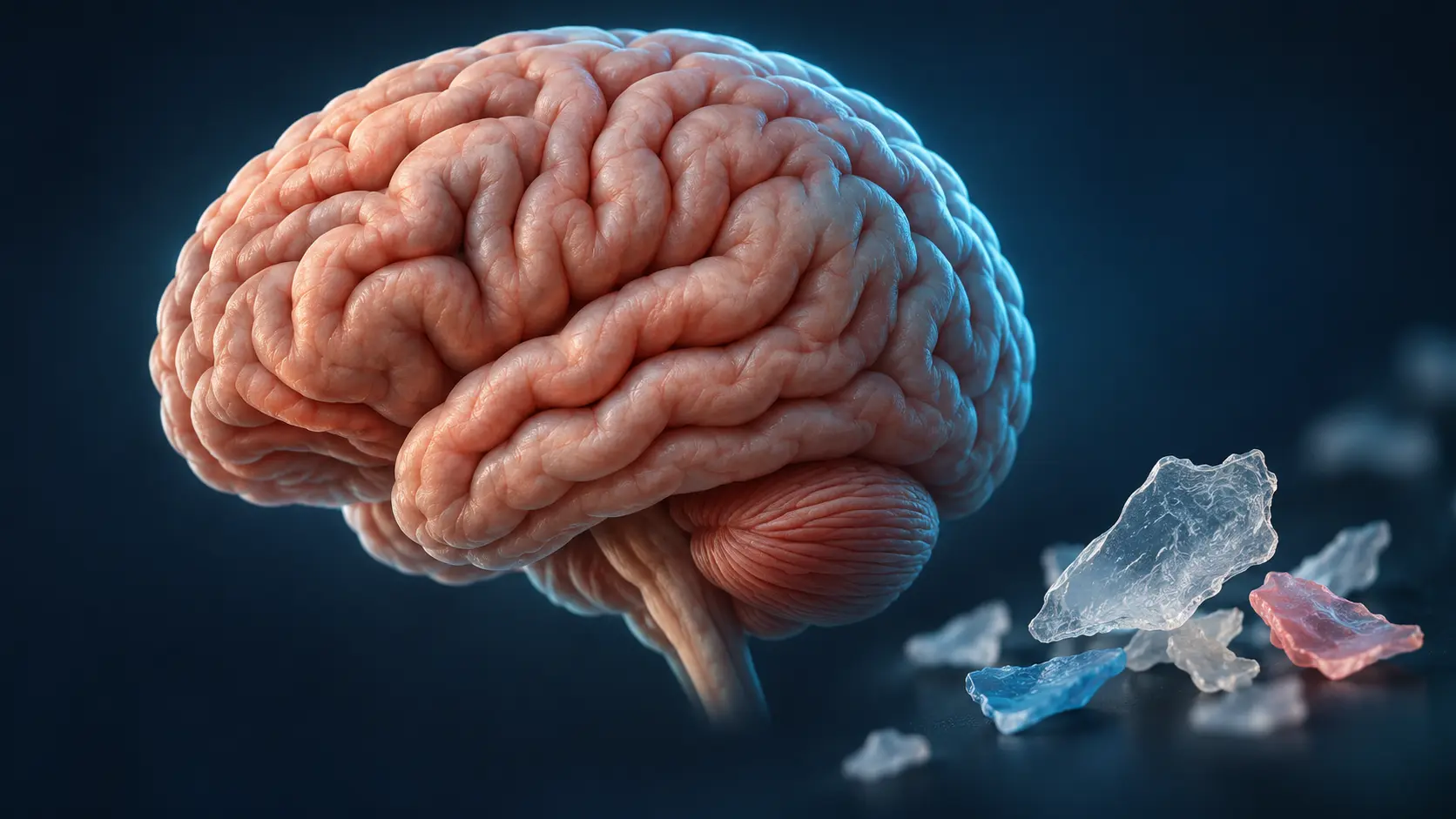
A 2025 study in Nature Medicine reported a median of 4.92 milligrams of micro- and nanoplastics per gram of frontal cortex tissue in human autopsy samples collected in 2024. In samples collected in 2016, the reported median was 3.35 milligrams per gram an increase of roughly 50% over eight years. These figures describe one region of the brain. The total amount in an average living person’s brain remains unknown.

How Much Plastic Did Researchers Find in Brain Tissue?

Researchers at the University of New Mexico examined tissue from the frontal cortex, a region near the front of the brain, working from autopsy specimens collected by the state medical investigator’s office. Each organ-and-year group included roughly 20–28 separate decedents.

Year samples were collected Reported median concentration Interquartile range Brain region sampled
2016 3,345 µg/g, or about 3.35 mg/g 1,267–5,213 µg/g Frontal cortex
2024 4,917 µg/g, or about 4.92 mg/g 4,026–5,608 µg/g Frontal cortex

The unit mg/g means milligrams of plastic per gram of tissue; 4,917 µg/g works out to plastic making up roughly 0.5% of the sampled tissue by weight. The median is the middle value for each group. The higher result in 2024 comes from comparing two groups sampled in different years the study did not track the same people over time.

Two details strengthen the time comparison. First, plastic concentrations showed no correlation with the decedent’s age (P = 0.87), sex, race/ethnicity, or cause of death the year of death was the factor that mattered. Second, the team also measured brain tissue collected between 1997 and 2013 from three East Coast brain banks; those samples had a lower median of 1,254 µg/g, consistent with rising concentrations over time, though geographic differences between the sample sources cannot be ruled out.

How Did Brains Compare With Other Organs?

Brain samples contained far more plastic than the liver and kidney samples from the same decedent groups. In 2024 samples, the median total plastic concentration was 433 µg/g in liver and 404 µg/g in kidney the brain median was roughly 10–12 times higher (P < 0.0001). Brain tissue also stood out for its composition: polyethylene made up about 75% of the plastic found in the brain on average, a larger share than in liver or kidney.

For context, the same team’s earlier work measured a median of 63.4 µg/g in human placentas, and other studies have reported around 299 µg/g in testes. Brain concentrations also resembled those reported in carotid artery plaques a comparison that matters because plaque buildup carrying microplastics has been linked to higher cardiovascular risk in earlier research.

Is There Really a Teaspoon of Plastic in the Brain?

Roughly yes by weight but only as an extrapolation, not a measurement. Applying the 2024 median of 4.92 mg/g to an average adult brain of about 1,300–1,400 g works out to roughly 6–7 grams of plastic about a teaspoon’s worth. The study never weighed a whole brain: researchers sampled the frontal cortex and, for the sake of the estimate, assumed the rest of the brain matches that concentration. No living person’s brain has been measured, and other regions could hold more plastic or less.

One caveat keeps the comparison honest: milligrams measure mass, while a teaspoon measures volume. A teaspoon holds about 5 grams of water, so “a teaspoon of plastic” is a weight comparison, not a literal spoonful and since polyethylene is lighter than water, an actual teaspoon of it would weigh slightly less.

How Did Scientists Measure Microplastics in Brain Tissue?

The team used a lab method called pyrolysis gas chromatography–mass spectrometry, or Py-GC/MS. It heats the processed sample and analyzes the chemicals released to estimate the mass of different plastics. Polyethylene was the most abundant plastic reported.

Two complementary methods supported the chemistry. Electron microscopy revealed that the isolated particles were largely nanoscale — shard- and flake-shaped fragments typically 100–200 nanometers long, which is why the paper’s figures cover both microplastics and nanoplastics. And an independent lab at Oklahoma State University re-analyzed a subset of the 2016 brain samples with Py-GC/MS and got consistent results, a check that the original measurements replicate across laboratories.

Why Is the Estimate Still Debated?

Measuring tiny plastic particles in human tissue is technically difficult, and in a follow-up commentary in Nature Medicine, Monikh and colleagues raised three main concerns about the study:

  • Contamination controls. The commentary argues the paper did not report the full set of quality-assurance procedures procedural blanks, field blanks, and validated digestion-and-recovery tests — recommended by international standards such as ISO 24187:2023, making it hard to rule out contamination from labware or airborne particles.
  • Lipid interference. Potassium hydroxide digestion, the step used to strip tissue away before analysis, is suboptimal for lipid-rich material. Brain tissue is about 60% lipid by dry weight (versus under 5% for liver and kidney), and incomplete digestion leaves residue behind.
  • False positives for polyethylene. Long-chain fatty acids release pyrolysis products that mimic polyethylene’s mass fragments. The commentary suggests the uniform PE dominance across organs may be an analytical artifact rather than a true exposure signature, and that the time trend could partly reflect differences in tissue lipid content.

The original team published a response defending its measurements: it points to the blank samples it did run, its screening of the polymer composition of every tube and pipette tip used, the independent replication by the second laboratory, and the fact that 2016 samples stored in plastic far longer than 2024 samples had lower concentrations, the opposite of what storage contamination would predict. The team also acknowledges method uncertainties in both directions, including a ~25% within-sample variability and a possible undercounting because ultracentrifugation may miss the smallest nanoparticles.

The 4.92 mg/g figure is the median reported by the study. Its precision, and how well it applies to other populations, remain open questions while methods are standardized.

What Did the Study Find About Dementia?

The researchers analyzed a separate group of 12 decedents with documented dementia (six Alzheimer’s disease, three vascular dementia, three other diagnoses) and reported a median concentration of 26,076 µg/g more than five times the 2024 normal-brain median. Under the microscope, dementia samples showed more plastic-like inclusions along blood vessel walls and in regions with accumulated immune cells.

Because the study examined tissue after death, it could not determine whether plastic exposure contributed to dementia, whether disease-related changes affected how much plastic was present, or whether other factors explained the difference. The authors note that brain atrophy, a leakier blood–brain barrier, and impaired clearance are all hallmarks of dementia that would be expected to raise measured concentrations no causality is assumed. The finding identifies a question for further research.

Key Takeaways

  • Brain frontal cortex samples held a median of 4.92 mg/g of plastics in 2024 — about 0.5% by weight and roughly 10–12× the levels in liver and kidney from the same year.
  • That is up ~50% from 2016 samples, with no link to age, sex, or cause of death.
  • The “teaspoon of plastic in your brain” headline is a whole-brain extrapolation of one region’s measurement — roughly 6–7 g by weight, not a measured total.
  • The methods and the exact figures are actively debated in the scientific literature; concentrations may be over- or underestimated.
  • A link to dementia was observed but is not evidence of causation.

Further Reading

Research sources

Technical Consultation

Need Help Applying Chitosan to Your Project?

Speak with our technical team about product selection, formulation, origin, molecular weight, DDA, samples, documentation, bulk pricing and commercial supply.

Product Selection Technical Guidance Sample & Bulk Support
Free Initial Discussion

Book a Consultation

Select a convenient time to discuss your application and purchasing requirements.

How Much Microplastic Is in the Human Brain?

How Much Microplastic Is in the Human Brain?

A 2025 study in Nature Medicine reported a median of 4.92 milligrams of micro- and nanoplastics per gram of frontal cortex tissue in human autopsy samples collected in 2024. In samples collected in 2016, the reported median was 3.35 milligrams per gram an increase of roughly 50% over eight years. These figures describe one region of the brain. The total amount in an average living person’s brain remains unknown.

How Much Plastic Did Researchers Find in Brain Tissue?

Researchers at the University of New Mexico examined tissue from the frontal cortex, a region near the front of the brain, working from autopsy specimens collected by the state medical investigator’s office. Each organ-and-year group included roughly 20–28 separate decedents.

Year samples were collected Reported median concentration Interquartile range Brain region sampled
2016 3,345 µg/g, or about 3.35 mg/g 1,267–5,213 µg/g Frontal cortex
2024 4,917 µg/g, or about 4.92 mg/g 4,026–5,608 µg/g Frontal cortex

The unit mg/g means milligrams of plastic per gram of tissue; 4,917 µg/g works out to plastic making up roughly 0.5% of the sampled tissue by weight. The median is the middle value for each group. The higher result in 2024 comes from comparing two groups sampled in different years the study did not track the same people over time.

Two details strengthen the time comparison. First, plastic concentrations showed no correlation with the decedent’s age (P = 0.87), sex, race/ethnicity, or cause of death the year of death was the factor that mattered. Second, the team also measured brain tissue collected between 1997 and 2013 from three East Coast brain banks; those samples had a lower median of 1,254 µg/g, consistent with rising concentrations over time, though geographic differences between the sample sources cannot be ruled out.

How Did Brains Compare With Other Organs?

Brain samples contained far more plastic than the liver and kidney samples from the same decedent groups. In 2024 samples, the median total plastic concentration was 433 µg/g in liver and 404 µg/g in kidney the brain median was roughly 10–12 times higher (P < 0.0001). Brain tissue also stood out for its composition: polyethylene made up about 75% of the plastic found in the brain on average, a larger share than in liver or kidney.

For context, the same team’s earlier work measured a median of 63.4 µg/g in human placentas, and other studies have reported around 299 µg/g in testes. Brain concentrations also resembled those reported in carotid artery plaques a comparison that matters because plaque buildup carrying microplastics has been linked to higher cardiovascular risk in earlier research.

Is There Really a Teaspoon of Plastic in the Brain?

Roughly yes by weight but only as an extrapolation, not a measurement. Applying the 2024 median of 4.92 mg/g to an average adult brain of about 1,300–1,400 g works out to roughly 6–7 grams of plastic about a teaspoon’s worth. The study never weighed a whole brain: researchers sampled the frontal cortex and, for the sake of the estimate, assumed the rest of the brain matches that concentration. No living person’s brain has been measured, and other regions could hold more plastic or less.

One caveat keeps the comparison honest: milligrams measure mass, while a teaspoon measures volume. A teaspoon holds about 5 grams of water, so “a teaspoon of plastic” is a weight comparison, not a literal spoonful and since polyethylene is lighter than water, an actual teaspoon of it would weigh slightly less.

How Did Scientists Measure Microplastics in Brain Tissue?

The team used a lab method called pyrolysis gas chromatography–mass spectrometry, or Py-GC/MS. It heats the processed sample and analyzes the chemicals released to estimate the mass of different plastics. Polyethylene was the most abundant plastic reported.

Two complementary methods supported the chemistry. Electron microscopy revealed that the isolated particles were largely nanoscale — shard- and flake-shaped fragments typically 100–200 nanometers long, which is why the paper’s figures cover both microplastics and nanoplastics. And an independent lab at Oklahoma State University re-analyzed a subset of the 2016 brain samples with Py-GC/MS and got consistent results, a check that the original measurements replicate across laboratories.

Why Is the Estimate Still Debated?

Measuring tiny plastic particles in human tissue is technically difficult, and in a follow-up commentary in Nature Medicine, Monikh and colleagues raised three main concerns about the study:

  • Contamination controls. The commentary argues the paper did not report the full set of quality-assurance procedures procedural blanks, field blanks, and validated digestion-and-recovery tests — recommended by international standards such as ISO 24187:2023, making it hard to rule out contamination from labware or airborne particles.
  • Lipid interference. Potassium hydroxide digestion, the step used to strip tissue away before analysis, is suboptimal for lipid-rich material. Brain tissue is about 60% lipid by dry weight (versus under 5% for liver and kidney), and incomplete digestion leaves residue behind.
  • False positives for polyethylene. Long-chain fatty acids release pyrolysis products that mimic polyethylene’s mass fragments. The commentary suggests the uniform PE dominance across organs may be an analytical artifact rather than a true exposure signature, and that the time trend could partly reflect differences in tissue lipid content.

The original team published a response defending its measurements: it points to the blank samples it did run, its screening of the polymer composition of every tube and pipette tip used, the independent replication by the second laboratory, and the fact that 2016 samples stored in plastic far longer than 2024 samples had lower concentrations, the opposite of what storage contamination would predict. The team also acknowledges method uncertainties in both directions, including a ~25% within-sample variability and a possible undercounting because ultracentrifugation may miss the smallest nanoparticles.

The 4.92 mg/g figure is the median reported by the study. Its precision, and how well it applies to other populations, remain open questions while methods are standardized.

What Did the Study Find About Dementia?

The researchers analyzed a separate group of 12 decedents with documented dementia (six Alzheimer’s disease, three vascular dementia, three other diagnoses) and reported a median concentration of 26,076 µg/g more than five times the 2024 normal-brain median. Under the microscope, dementia samples showed more plastic-like inclusions along blood vessel walls and in regions with accumulated immune cells.

Because the study examined tissue after death, it could not determine whether plastic exposure contributed to dementia, whether disease-related changes affected how much plastic was present, or whether other factors explained the difference. The authors note that brain atrophy, a leakier blood–brain barrier, and impaired clearance are all hallmarks of dementia that would be expected to raise measured concentrations no causality is assumed. The finding identifies a question for further research.

Key Takeaways

  • Brain frontal cortex samples held a median of 4.92 mg/g of plastics in 2024 — about 0.5% by weight and roughly 10–12× the levels in liver and kidney from the same year.
  • That is up ~50% from 2016 samples, with no link to age, sex, or cause of death.
  • The “teaspoon of plastic in your brain” headline is a whole-brain extrapolation of one region’s measurement — roughly 6–7 g by weight, not a measured total.
  • The methods and the exact figures are actively debated in the scientific literature; concentrations may be over- or underestimated.
  • A link to dementia was observed but is not evidence of causation.

Further Reading

Research sources

Recent Posts

Access this document

Please enter your details to download the file.
Thank you. You can now access the document below.